When In Doubt, Get the Dermatoscope Out!
NO
PHARMACEUTICAL INFLUENCE
When In Doubt, Get the Dermatoscope Out!

John is one of your regular patients, a 64-year-old stoical tree surgeon, who presents with a new pigmented papule on his torso. For someone normally unphased by challenging work environments, this has him completely flummoxed. “I’ve researched this online and since reading the possible diagnosis, I’ve started to write a Will, I’m actually on my way to hand in my notice…” he confesses.
After confirming this is a new slowly evolving asymptomatic lesion present on John’s back, you feel like Sherlock Holmes (your dermatoscope replacing the magnifying glass) and as you start your inspection … et voilà! Views on dermoscopy reveal multiple comedo-like openings, milia-like cysts and that classic sharp demarcation at the periphery, not to mention those ‘fat fingers’ [this is a real dermoscopy term!] and with a sigh of relief you reassure John this is a benign seborrhoeic keratosis. His relief is palpable, and with a slightly sheepish explanation that he was of course ‘joking’ about his job resignation, he’s on his way, one very happy chappy!
Compare this to your next case, Tina, a 50-year-old beautician who admits to occasionally using the salon’s sun-bed from time-to-time. She presents with an evolving pigmented nodular lesion on her thigh, reaching for your faithful side-kick, your dermatoscope, this reveals streaks at the periphery, an atypical pigment network, a blue-white veil, not to mention those polymorphous vessels, after your prompt 2-week-wait referral, Tina is diagnosed with a 2.5mm Breslow thickness nodular melanoma, and thanks to your expert diagnostic skills, this is now completely excised.
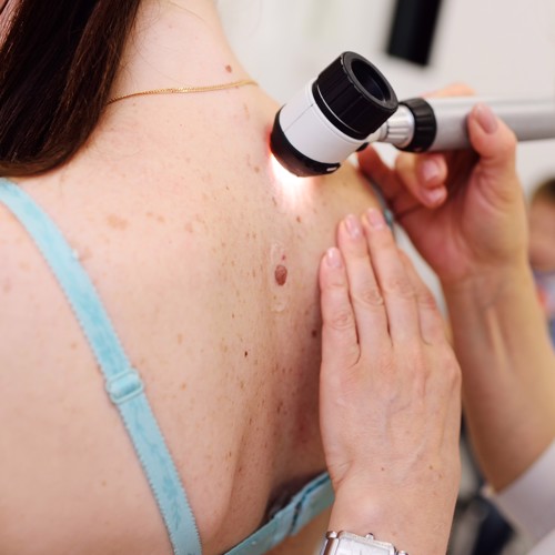
So how does dermoscopy work? The dermatoscope is not just an illuminated magnifying glass (although it can be used as one), but it also lets you see through the surface of the skin to the structures that lie beneath it.
How can dermoscopy help us in primary care? Well, we’ve saved John’s job for a start! On a slightly more serious note, dermoscopy can be invaluable for pigmented and non-pigmented skin lesions; rashes; hair, scalp and nail problems; head lice (and it is now the gold standard diagnostic test for scabies) – and even for telling real diamonds from fakes! For those of us who have a dermatoscope at our practice, being able to utilise this to its full advantage allows us to send high quality images through to our dermatology colleagues to improve the advice and guidance received.
If you’d like to learn more about the magic of dermoscopy, including practical tips on using the dermatoscope, how to optimise the photo quality and an overview of the dermoscopic features of common skin lesions seen in primary care, please join our FREE interactive one-hour Introduction to Dermoscopy Webinar on Tuesday 7th November at 8pm, or watch on demand, where Dr Will Duffin and I are joined by the incredibly talented Dr Chin Whybrew (GP Partner, dermoscopy lead for the Primary Care Dermatology Society and freelance dermatology and dermoscopy tutor for Cardiff University), where we will all share our words of wisdom and passion for dermoscopy with you. We look forward to seeing you then!
Dr Philippa Davies
27th September 2023
Join us at one of our upcoming courses or live webinars

Recent NB Blogs
Need an immediate update? – all our courses are available on demand
Did you find this useful?
You can quickly add CPD to your account by writing a reflective note about the When In Doubt, Get the Dermatoscope Out! post you've read.
Log in to your NB Dashboard and use the 'Add Reflective Note' button at the bottom of a blog entry to add your note.

